Have you ever wondered what fruits and vegetables might look like if they were scanned underneath an MRI? Yes? Well, look no further! A project by Andy Ellison, an MRI technologist who works at the Boston University Medical School, reveals fascinating images of pears, bananas, and more under the machine.
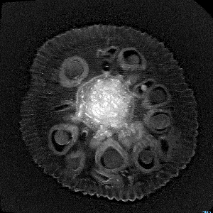
The Magnetic Resonance Imager (MRI) uses magnets to align hydrogen atoms and radio waves to produce an image. When done in layered succession, a three-dimensional image of the item being examined is produced. And, after scrolling through the collection below, we think you’ll agree that the result is amazing. The lines and shapes of the fruits and vegetables resemble veins and capillaries, similar in appearance to living organisms.
Learn more by visiting Andy’s website here.
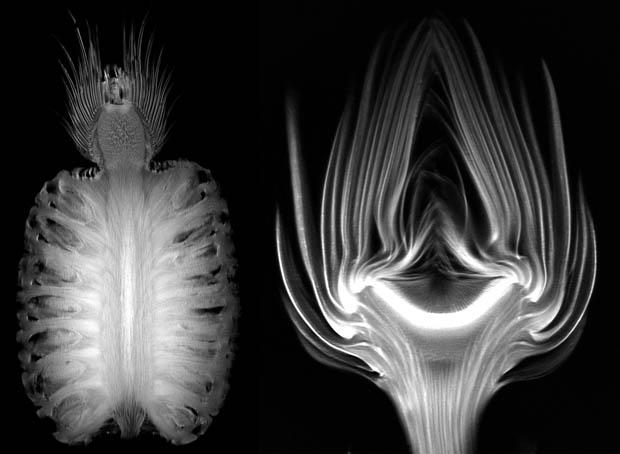
#1 Pear
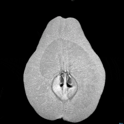
#2 Banana
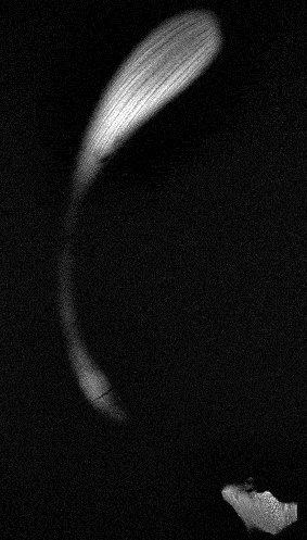
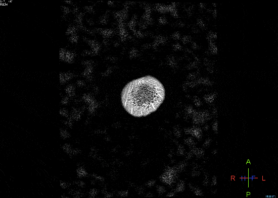
#3 Garlic
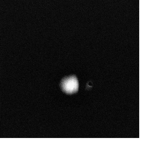
#4 Jackfruit
#5 Pineapple
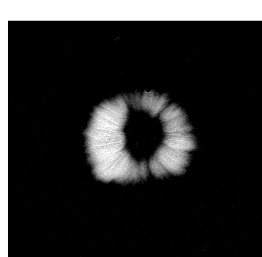
#6 Onion
#7 Passion Fruit
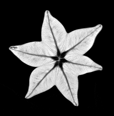
#8 Starfruit
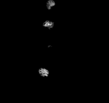
#9 Strawberries
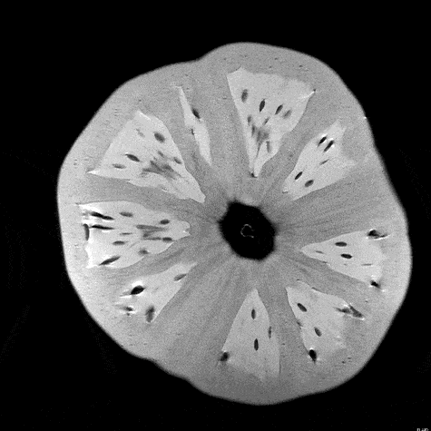
#10 Tomato
#11 Pumpkin
#12 Watermelon
What are your thoughts? Please comment below and share this news!
This article (What Happens When Fruits And Vegetables Are Scanned Under An MRI Is Amazing) is free and open source. You have permission to republish this article under a Creative Commons license with attribution to the author and TrueActivist.com